The b -globin gene family
The human genome consists of approximately 3 billion base pairs. Until recently, scientists had predicted that this large amount of DNA would encode almost 100,000 different genes; however, now that the sequencing of the entire human genome is almost complete, that number has dropped to only 30,000 genes. In fact, close analysis of the genome has shown that well over 90 percent of the genome consists of non-functional DNA.
Of the remaining functional DNA, 25-50 percent of the genes encoding proteins are found only once in the haploid genome and are known as solitary genes. Frequently, the DNA surrounding a particular gene contains sequences that are close, but not identical, copies of the gene. These multiple copies are believed to be the result of duplication and divergence. This is the process by which a single gene is first duplicated and then undergoes selective pressure to mutate into a gene that is similar, but not identical, in sequence to its ancestral gene. One such example of this process is the development of the beta-globin gene family. After globin family genes from different species were sequenced, an evolutionary tree predicting the development of the globin family of genes was developed. A proposed tree is shown below and illustrates the divergence of genes from leghemoglobin in plants to hemoglobin and myoglobin in animals. At each branch point the gene has duplicated and then mutates into a new, but similar gene. For example, at some point in history the common ancestral gene first underwent a duplication. The two copies that resulted then mutated in different ways; one forming leghemoglobin another forming insect hemoglobin.
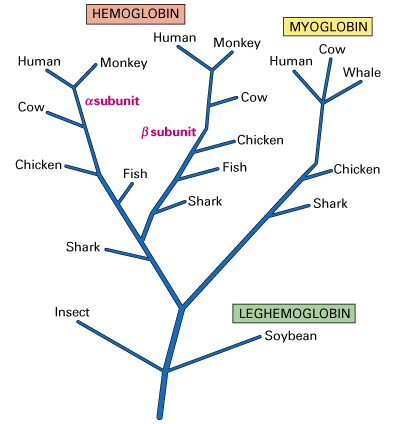
Figure 1: Diagram of Globin Family Tree (Lodish et.al.,2000)
A set of duplicated genes that encode proteins with similar but nonidentical amino acid sequences is known as a gene family (Lodish et.al., 2000). Numerous different gene families have been identified with various functions. For example, the formation of a functional hemoglobin molecule requires the use of products from two such gene families by combining two b -globin family polypeptides with two a -globin famlily polypeptides and four small heme groups. The beta-globin gene family located on chromosome 11 is shown in the figure below and consists of five functional genes (blue boxes) and two pseudogenes (diagonal lines).
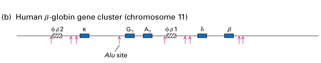
Figure 2: The Beta-globin Gene Family on Chromosome 11 (Lodish et.al., 2000)
All of the hemoglobins encoded by these different genes function to carry oxygen in the blood; however, each gene exhibits specific variations in function. For example, the epsilon globin gene is normally expressed in the embryonic yolk sac while the Ag and Gg genes are expressed only during fetal development. These hemoglobin proteins have a higher binding affinity for oxygen than the adult hemoglobins encoded by the b and d genes. This increased binding allows the fetus to successfully extract oxygen from the blood without competing with the mother. Adult hemoglobin has a lower oxygen affinity allowing better release of oxygen to tissue, especially muscle.
Two regions of the globin gene family contain nonfunctional sequences known as pseudogenes (diagonal lined boxes). These genes are similar to their functional globin gene counterparts but are no longer transcribed into mRNA because of changes in sequence that have occurred during the course of their evolution.
Even a slight change in one of the genes encoding a subunit of the hemoglobin molecule can have disastrous results. This laboratory will focus on two diseases that result from different mutations in the beta-globin gene.
Click on the link below to learn more about Sickle Cell Anemia.