Sickle cell anemia occurs in 1 in 500 individuals of African descent and is characterized by red blood cells that are rigid and sickled in shape (see Figure 3).
Figure 3: Comparison of healthy cells on left with sickled cells on right. (Campbell, 2000)
The difficulty these sickle cells have in moving through the circulatory system has numerous effects on those who suffer from the disease including anemia, brain damage, and kidney failure (effects are further illustrated in Figure 4).
Figure 4: Diagram of Physiological Effects of Sickle Cell Anemia(Campbell, 2000)
Believe or not, all of the symptoms listed in the figure above are caused by a single point mutation in codon six of the b -globin gene from glutamic acid to valine (See figure below). This small difference produces a mutant protein that is insoluble in the red blood cell, and consequently, forms a crystalline structure.
Figure 5: a) Amino acid sequence of functional hemoglobin with glutamic acid in position 6, b) Amino acid sequence for sickled hemoglobin with valine in position 6. (Campbell, 2000)
There are, however, advantages to carrying only one copy of the mutant b -globin gene. These heterozygote individuals do not suffer from the disease and exhibit increased resistance to malaria because the parasite which causes the disease is unable to reproduce in the red blood cells of heterozygote individuals. Malaria occurs most frequently in tropical regions near the equator which helps to explain why those of African descent are more commonly affected.
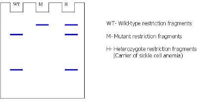
Considering the devastating effects of sickle cell anemia, parents may want to determine if they are carriers of the disease before they decide to have children. If two carriers mate, 25 percent of their children will be homozygous for the mutant gene. The use of restriction enzymes now enables carriers to be determined by examining RFLPs, or restriction fragment length polymorphisms. Mutations in DNA that add or delete a restrictions site can be determined by analysis using gel electrophoresis after digesting DNA with a restriction enzyme. First compare the restriction sites (shown in blue) found on the wild-type and mutant b -globin genes:
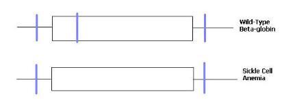
Notice that the point mutation in codon six eliminates one of the restriction sites normally found in the wild-type copy of the b -globin gene. If these two genes were digested with restriction enzyme and run out on a gel you would expect the following results:
Your instructor will use sickle cell anemia to illustrate how to use the various databases in this laboratory exercise. Click on the link below to return to the Bioinformatics Laboratory Homepage.